Beyond Fibroblasts: CHPs provide Key Evidence for Keratinocyte-Driven Skin Development

Despite collagen being the most abundant protein in the skin, the process of how the dermal collagen lattice forms isn't completely understood. In large part, this is because mammalian skin is opaque, limiting the options researchers have to observe collagen formation in living tissue. To overcome this, a groundbreaking study is using axolotls - which have transparent skin - to shed light on this fundamental process. Not only does this research change our understanding of skin biology - It's also paving the way for advancements in cosmetics and skin medicine.
This study challenges the long-held belief that dermal fibroblasts that are solely responsible for producing Type I Collagen , the main component of the skin's dermis. Instead this research found that keratinocytes also significantly contribute to dermal collagen formation, especially in early stakes of dermis development. One key method used to show keratinocyte contributions was an in-vivo "pulse-chase" experiment.
Read More About the Study's Findings
A Pulse-chase experiment involves the sequential subcutaneous injections of spectrally-distinct fluorescent collagen probes, DAR followed by DAF, allowing visualization of existing versus newly synthesized collagen.

In early axolotl developmental stages, confocal microscopy revealed that the DAF (chase) signal, representing nascent collagen, localized to the predominantly to the apical side of the developing dermis, subjacent to the epidermal-dermal junction. The DAR (pulse) signal, showing pre-existing collagen, was distributed more evenly. This spatial deposition pattern, new collagen being developed from the dermal-epidermal junction downwards, strongly implicates keratinocytes as a primary source of secreted collagen during this phase of development.
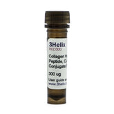
To further validate their findings and assess the conservation of this mechanism, researchers utilized Collagen Hybridizing Peptides (CHPs) in a complementary pulse-chase experiment performed in zebrafish. CHPs are 3Helix's proprietary technology, engineered to selectively bind to damaged / remodeling collagen molecules, offering an extremely precise method for labeling sites of active collagen turnover and deposition.

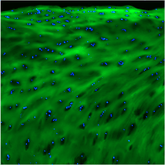
In their zebrafish model, larvae were initially stained with DAF in-vivo, 12 days post-fertilization (dpf). Following 5 days of growth, the fixed larvae were stained with R-CHP, Collagen Hybridizing Peptides fluorescently labeled with Cy3 dye. Consistent with the axolotl observations, the R-CHP signal exhibited preferential accumulation towards the apical side, in line with their earlier pulse-chase experiment in axolotls. This independent line of evidence, obtained using CHPs in a different vertebrate model, corroborates the axolotl data and suggests that keratinocyte-mediated collagen deposition is an evolutionarily-conserved process during vertebrate skin development. Further support came from detecting procollagen protein within epidermal cells in both models.
The study by Ohashi et al. provides significant evidence revising the canonical view of dermal collagen synthesis. By demonstrating a substantial contribution from keratinocytes in establishing the initial dermal matrix, the research underscores a more complex interplay between epidermal and mesenchymal cells than previously recognized.
The application of advanced imaging techniques, including sequential fluorescent probe labeling and the use of CHPs, proved instrumental in visualizing these cellular dynamics in vivo and in histology, challenging established models. These findings not only advance our fundamental understanding of skin development but also hold implications for regenerative medicine. A refined model of dermal collagen formation, acknowledging the role of keratinocytes and elucidated by tools like CHPs, may inform novel approaches for promoting functional skin regeneration and developing therapies for fibrotic skin diseases or age-related dermal changes.
Reference:
|
Ohashi, A., Sakamoto, H., Kuroda, J. et al. Keratinocyte-driven dermal collagen formation in the axolotl skin. Nat Commun 16, 1757 (2025). https://doi.org/10.1038/s41467-025-57055-7
|
|
