Delayed Fracture Healing in Obesity-associated Type 2 Diabetic Mouse Model
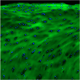
In this interesting article published in Bone, researchers from Penn State utilized CHPs to help monitor fracture healing in the diet-induced obesity (DIO) mouse model. They applied numerous characterization techniques to probe the microstructure of collagen within a wound (fracture) healing environment. They evaluated collagen structure in a healthy (lean) mouse model and compared it with the DIO model using second harmonic generation (SHG), micro-CT, as well as immunofluorescent staining (IF) to investigate how obesity influences the bone healing response at a fracture site. They found significantly higher levels of CHP signal in the DIO callus than the lean callus, indicating there was a high content of damaged collagen (Figure 7).. This result corroborated well with their other study involving advanced glycation end products (AGEs) which showed a higher accumulation of AGEs in the fracture site of the DIO model (Figure 8). This is important because AGEs inhibit ECM remodeling by interfering with collagen degradation, leading to pathological accumulation of partially degraded collagen fibers. They are the first group to thoroughly investigate the impact of the structure of fibrillar collagen has on the healing process.

Fig. 7. DIO callus has elevated levels of unfolded collagen helices. (A) Representative images of fluorescent staining (green) with collagen hybridizing peptide (CHP) that binds only to denatured, unfolded collagen strands. Staining was performed at the indicated post-fracture time points. Scale bar = 100 μm. (B) Quantitation of CHP-stained area normalized to total Col I area. Analysis was performed in 6 lean and 6 DIO callus tissues; 3 sections were analyzed in each. Bar graphs represent the average ± SEM. (*) P < 0.05 using student ttest and time-matched lean callus as a control. (For interpretation of the references to colour in this figure legend, the reader is referred to the web version of this article.)
ABSTRACT: Impaired fracture healing in patients with obesity-associated type 2 diabetes (T2D) is a significant unmet clinical problem that affects millions of people worldwide. However, the underlying causes are poorly understood. Additionally, limited clinical information is available on how pre-diabetic hyperglycemia in obese individuals impacts bone healing. Here, we use the diet-induced obesity (DIO) mouse (C57BL/6J) model to study the impact of obesity-associated pre-diabetic hyperglycemia on bone healing and fibrillar collagen organization as healing proceeds from one phase to another. We show that DIO mice exhibit defective healing characterized by reduced bone mineral density, bone volume, and bone volume density. Differences in the healing pattern between lean and DIO mice occur early in the healing process as evidenced by faster resorption of the fibrocartilaginous callus in DIO mice. However, the major differences between lean and DIO mice occur during the later phases of endochondral ossification and bone remodeling. Comprehensive analyses of fibrillar collagen microstructure and expression pattern during these phases, using a set of complementary techniques that include histomorphometry, immunofluorescence staining, and second harmonic generation microscopy, demonstrate significant defects in DIO mice. Defects include strikingly sparse and disorganized collagen fibers, as well as pathological accumulation of unfolded collagen triple helices. We also demonstrate that DIO-associated changes in fibrillar collagen structure are attributable, at least in part, to the accumulation of advanced glycation end products, which increase the collagen-fiber crosslink density. These major changes impair fibrillar collagens functions, culminating in defective callus mineralization, remodeling, and strength. Our data extend the understanding of mechanisms by which obesity and its associated hyperglycemia impair fracture healing and underline defective fibrillar collagen microstructure as a novel and important contributor.
Continue reading the FULL ARTICLE HERE!
