Can Physical Cues Prevent Scarring? Using CHPs to Investigate Myofibroblast Behavior
The link between tissue architecture and function is pivotal for the human cornea to properly function, since a highly organized collagen matrix is essential for optical transparency and vision. Following an injury to the cornea, aberrant healing results in a disorganized matrix which impedes light transmission through the cornea and leads to vision loss.
This process has raised a key question for researchers at the University of Edinburgh: can tissue-remodeling cells be guided to regenerate an organized, functional extracellular matrix (ECM) based on physical cues alone? To investigate, these researchers developed 3-D models of organized (aligned) and disorganized (random) corneal tissue using TGFB-1 stimulated corneal myofibroblasts suspended in a fibrinogen-based hydrogel. Comparing the two systems as models of healthy and fibrotic matrices provides insights into the role matrix alignment plays on corneal healing.

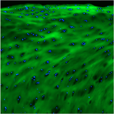
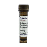
To aid their investigations, the researchers utilized Collagen Hybridizing Peptides (R-CHP, 3Helix) throughout their study. By selectively binding to denatured and remodeling collagen molecules, CHPs offered a clear view of matrix organization and collagen damage. This was first useful when researchers sought to differentiate the models they created. When examining their aligned and random models, they noted an aligned pattern of denatured collagen in their aligned constructs, and no discernable pattern in their random constructs.
Continue Reading
Beyond confirming the physical architecture, the CHP data provided crucial context for interpreting the biochemical composition of the newly formed matrix. With the structural differences validated, researchers confidently linked the tissue’s organization to its protein expression. They discovered that myofibroblasts in their aligned constructs produced significantly less scar-associated collagen I than their random constructs. Furthermore, fibronectin synthesis (a protein critical for ECM assembly) was enhanced in the aligned group, and CHP imaging revealed that fibronectin was highly colocalized with the newly formed collagen network. These data together allowed the researchers to conclude that a healthier physical structure is directly associated with a more functionally regenerative ECM composition.

In conclusion, this research demonstrates that physical cues from the microenvironment are powerful regulators of myofibroblast behavior. The study showed that even when cells are chemically stimulated to form scars, an organized matrix can guide them to produce a more regenerative and less fibrotic ECM. Collagen Hybridizing Peptides were instrumental in proving this connection, linking the matrix's physical structure to the matrix composition profile. These findings open new avenues for developing anti-fibrotic therapies, suggesting that future treatments could focus on physically guiding tissue regeneration to prevent scarring.
Paper: https://tvst.arvojournals.org/article.aspx?Giannopoulos, Antonios, Ludvig J. Backman, and Patrik Danielson. "Tissue Architecture Modulates Compositional and Structural Properties of Corneal Myofibroblast-Derived Matrix." Translational Vision Science & Technology 14.9 (2025): 9-9.
