First Collagen Hybridizing Peptide Application for Collagen Damage to Nervous Tissue: CHPs Help Confirm Distinct Patterns of Structural Damage in Epineuroclasis and Endoneuroclasis.
Peripheral Nerve Injuries (PNIs) represent a significant clinical challenge, frequently resulting in disability and chronic pain, particularly in younger patients. Recovery following PNI is oftentimes unpredictable and the decision to pursue surgical intervention is complicated by a limited ability to accurately assess the severity and extent of tissue damage.
The current PNI classification system, The Seddon/Sunderland system (Introduced by Seddon to be later improved upon by Sunderland), describes an “inside-out” cascade of structural failure, wherein the innermost structures of the endoneurium fail first, progressing outwards towards the epineurium. While the Seddon/Sunderland system has been widely used for decades, the field has yet to reliably detect and differentiate the specific injury grades defined by this classification. Furthermore, various animal models have struggled to replicate the specific structural changes proposed by Seddon & Sunderland. These clear discrepancies indicate that there are significant limitations in our understanding of the mechanisms underlying peripheral nerve injuries.
Continue Reading
Considering the frequency peripheral nerve injuries occur, the unknowns surrounding the recovery process, and the life-altering consequences of inadequate treatment, there is a pressing need for an improved understanding of PNI mechanics and for the development of improved diagnostic tools which accurately assess structural damage.
A study just published in Clinical Orthopedics and Related Research, Schröen et al. (2025), introduced a novel approach to investigate structural damage in a rat model of median nerve stretch injury.

This innovative study utilized Collagen Hybridizing Peptides (CHPs) to visualize and characterize collagen damage, providing new insights into the sequence of mechanical and structural failure during nerve stretch. This is the first time in literature a study has used CHPs to stain nervous tissue for mechanical damage.
Specifically, Schröen et al. (2025) aimed to:
- Define the pattern of mechanical failure during a nerve stretch injury
- Examine the sequence of structural degradation of specific anatomical substructures, including collagen, during failure.
Collagen Hybridzing Peptides selectively bind to “damaged collagen”: collagen molecules that have undergone structural alterations (denaturation, remodeling, etc.) that disrupts their native triple-helical conformation. In the study, CHPs were employed to specifically identify collagen molecules mechanically damaged during nerve stretch injury experiments. Like most tissues, collagen is a crucial structural protein in peripheral nerves. Specifically, collagen is a key component of the:
- Epineurium: The outermost layer surrounding the entire nerve, providing protection and structural integrity
- Perineurium: A protective sheath surrounding nerve fascicles
- Endoneurium: The delicate connective tissue surrounding axons.
Detecting damaged collagen in these structures serves as a valuable marker of structural injury in response to mechanical stress. In the case of Schröen et al. (2025), CHPs specifically pinpointed the location and extent of collagen damage resulting from nerve stretch, providing insights into injury mechanisms.
The study by Schröen et al. used an in vivo rat model to study peripheral nerve stretch injuries, employing a load-to-failure experiment paired with electrical nerve stimulation, histology, and immunohistochemistry. This comprehensive approach allowed them to clearly define the sequence of mechanical and structural failure in rat medial nerves. The study’s load-to-failure experiments revealed two distinct mechanical failure events, which they labeled epineuroclasis followed by endoneuroclasis. These events were identified on load-deformation curves (below).

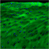
CHPs were crucial for visualizing the structural damage associated with each event. At epineuroclasis, CHPs revealed moderate collagen damage which accompanied the disruption of the endoneurium, axonal damage, and hemorrhage. At Endoneuroclasis, CHPs showed extensive molecular collagen damage throughout the endoneurium, with the greatest damage occurring at the site of initial epineurium rupture.

This clearly demonstrated the progressive nature of collagen damage, starting at the epineurium and then extending inward. This represents a key finding, contradicting the “inside-out” view proposed by Sunderland. A better, comprehensive understanding of mechanical failure progression in peripheral nerve injury has great implications for developing more accurate diagnostic tools, prognoses, and improving treatment strategies. By providing a more detailed understanding of the sequence of structural failure, Schröen et al. (2025) have not only advanced our fundamental knowledge of nerve injury mechanisms but also paves the way for future research aimed at improving outcomes for patients with peripheral nerve injuries.
Full Paper:
Schroen, Christoph A. BS1,2,3; Duey, Akiro H. BS1; Nasser, Philip MS1; Laudier, Damien BS1; Cagle, Paul J. MD1; Hausman, Michael R. MD1. What Is the Sequence of Mechanical and Structural Failure During Stretch Injury in the Rat Median Nerve? The Neuroclasis Classification. Clinical Orthopaedics and Related Research ():10.1097/CORR.0000000000003405, February 18, 2025. | DOI: 10.1097/CORR.0000000000003405
