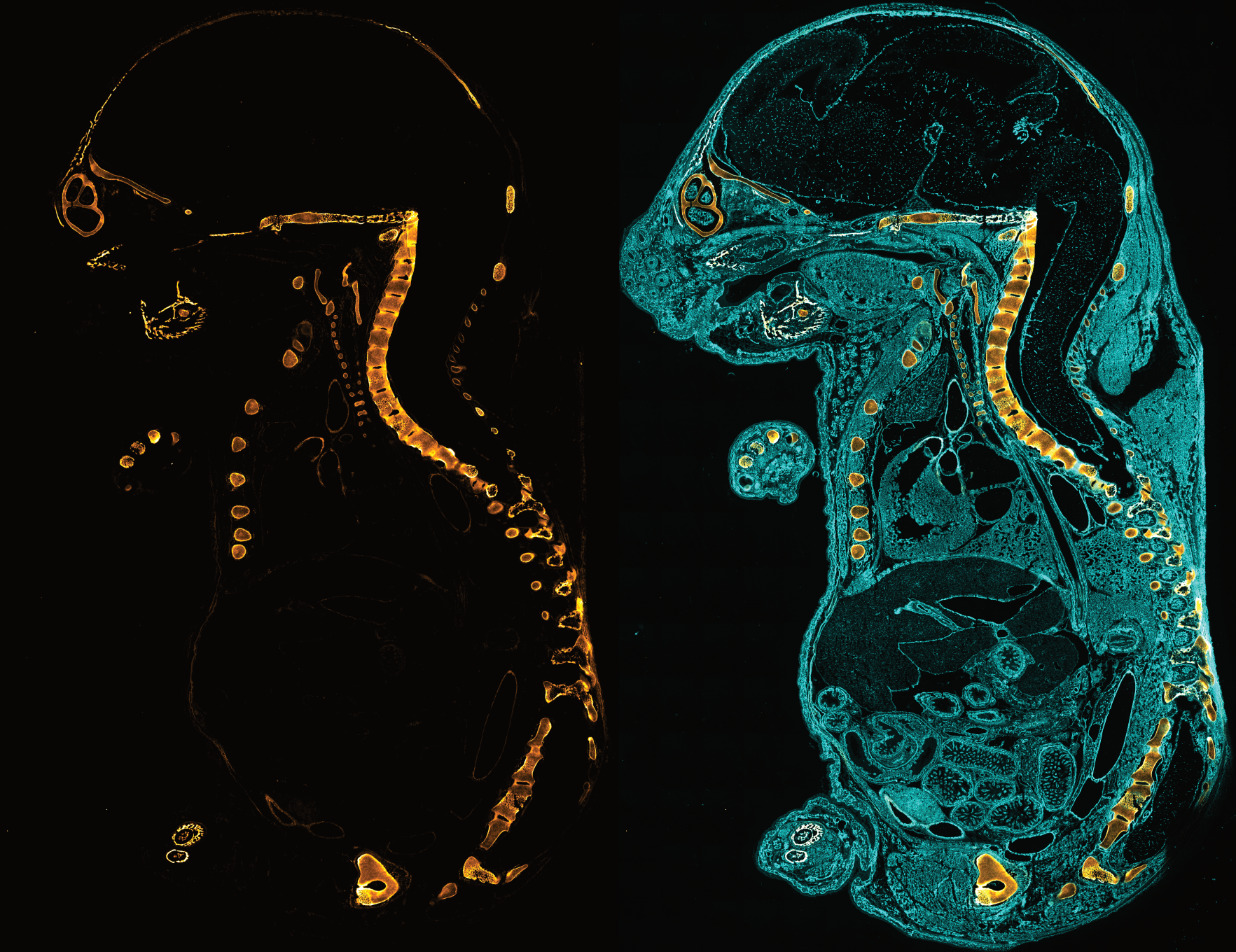
Collagen is the major building block in all load bearing tissues including tendon, ligament, bone, and cartilage. Mechanical injury to these tissues causes changes in collagen structure which can initiate pathological processes without detectable morphological changes at the macroscale.
Utilizing the collagen hybridizing peptide (CHP), which binds to the unfolded collagen chains by triple helix formation, researchers can now detect molecular level subfailure damage to collagen in mechanically stretched tendon fascicles.[1] The results directly reveal that the collagen triple helix unfolds as a result of tensile loading, and that the irreversible unfolding of collagen molecules can accumulate in tendon fascicles under cyclic fatigue.[1]

Left: Fluorescence scans of rat tail tendon fascicles that have been uniaxially loaded to 5, 10.5, and 15% strain for a single cycle and stained by 5-FAM-labeled CHP (F-CHP), clearly showing an increase of CHP staining with increased strain but minimal fluorescence signal outside the stretched region. A corresponding photo of the 15% strain sample is provided. Scale bars: 2 mm.
Right: CHP intensity quantified from fluorescence images of loaded fascicles (green) begins to increase from baseline as the stress-strain curve (black) deviates from linearity (red line), suggesting that the unfolding of collagen molecules corresponds with the initiation of the mechanical damage.

Left: Multiphoton images of collagen SHG (gray) and fluorescence from CHP binding (green) in a 15% strained rat tail tendon fascicle. CHP staining pattern revealed heterogeneity in molecular level collagen damage. Scale bars: 100 μm.
Right: A TEM image of CHP-conjugated gold nanoparticles binding to damaged collagen fibrils in a 12% strain fascicle, exhibiting a high density of nanoparticle in areas with visible fibril disruption (loss of d-banding pattern). Scale bar: 200 nm. A similar experiment can be conducted in your study by incubating the fibrils with our biotin-labeled CHP (B-CHP) followed by visualization with TEM using commercial streptavidin-conjugated gold nanoparticles.
The CHP probes open up a new avenue for detection and quantitative comparison of mechanical damage to collagen in all connective tissues, including bone, cartilage, tendon, ligament, intervertebral discs,[2] heart valves, blood vessels,[3] skin, cornea[4] and etc., allowing understanding of the mechanical behaviors and damage mechanisms of these tissues at the molecular level.


